![]()
R Banding by Fluorescence Using Acridine Orange (AO)
![]()
|
Principle R-banding
methods are useful for analyzing deletions or translocations that involve the
telomeres of chromosomes. Background Acridine
orange was originally used to stain untreated chromosomes, both human and
mouse. Bobrow et al. and Baserga and Castoldi independently reported the use
of acridine orange to obtain a reverse banding pattern of chromosomes.
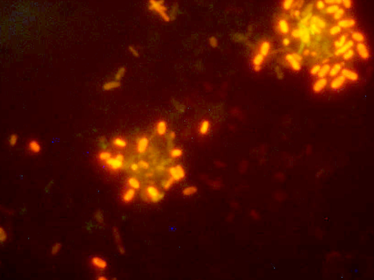
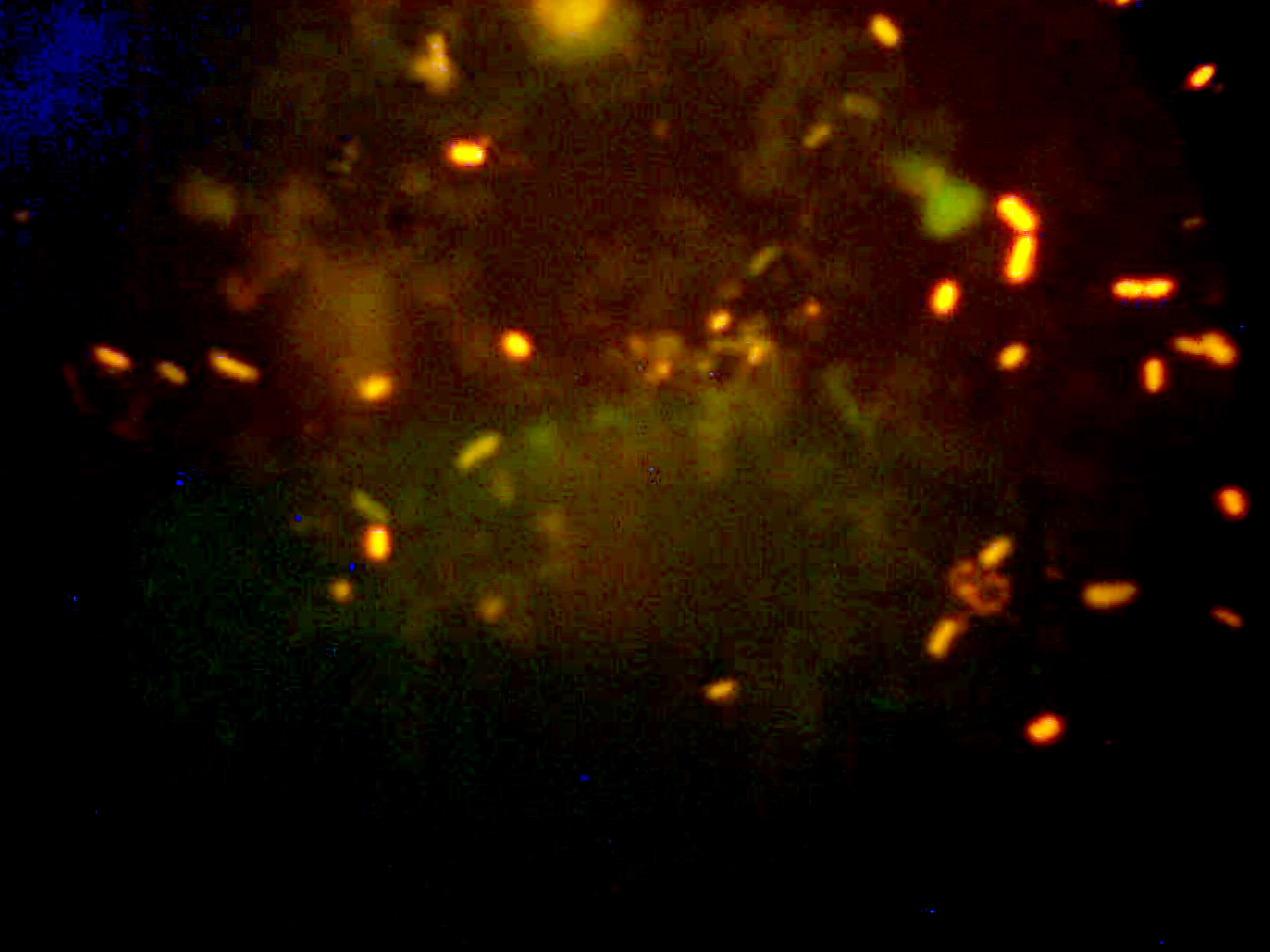
Acridine orange (AO) is a base composition-independent fluorochrome that
binds to DNA by intercalation and which gives relatively uniform fluorescence
along the length of the chromosome arms. The dye binds very little to
non-nucleic acid cell components, but it fluoresces orange-red when bound to
single-stranded nucleic acids and yellow-green when bound to double-stranded
nucleic acids. Following hot phosphate buffer treatment, R bands are
yellow-green, and G/Q bands are orange-red. The major factor that contributes
to R banding is the relative GC-richness of the R bands. In many
laboratories, RHG methods have been abandoned in favor of a fluorescent
R-banding technique (Gustashaw, 1991). Solutions
32 ml of
0.07N Na2HPO4 . 12 H20 68 mL of
0.07 mol/L KH2PO4 Adjust pH to 6.5 by adding 0.07N Na2HPO4 . 12 H2O to the solution.
Procedure
Click Here for Image Click Here for Image
References Gustashaw,
KM. Chromosome Stains. In The ACT
Cytogenetics Laboratory Manual, Second Edition, edited by M. J. Barch.
The Association of Cytogenetic Technologists, Raven Press, Ltd., New York,
1991. Verma,
RS, Lubs HA. Additional observations on the preparation of R banded human
chromosomes with acridine orange. Can J Genet Cytol 18:45-50, 1976. Verma,
RS, Lubs HA. A simple R banding technique. Am J Hum Genet 27:110-117, 1975. |
|
|